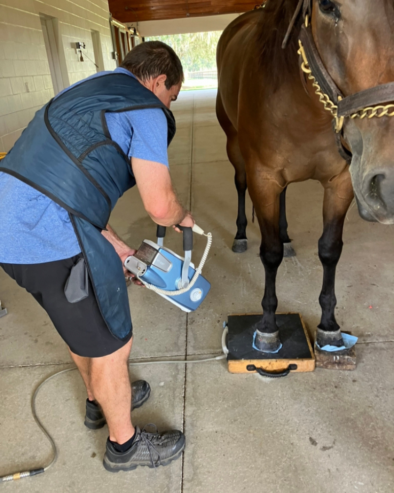
Dr. Barrett doing dinosaur radiographs!

Digital radiographs are mainly used to evaluate bone conditions such as arthritis, fractures, tendon calcifications, infections, and to measure foot balance.
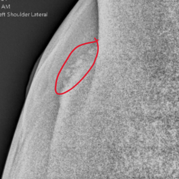
Radiograph of the scapular spine showing fragments from a kick to the shoulder.

Scapular Spine

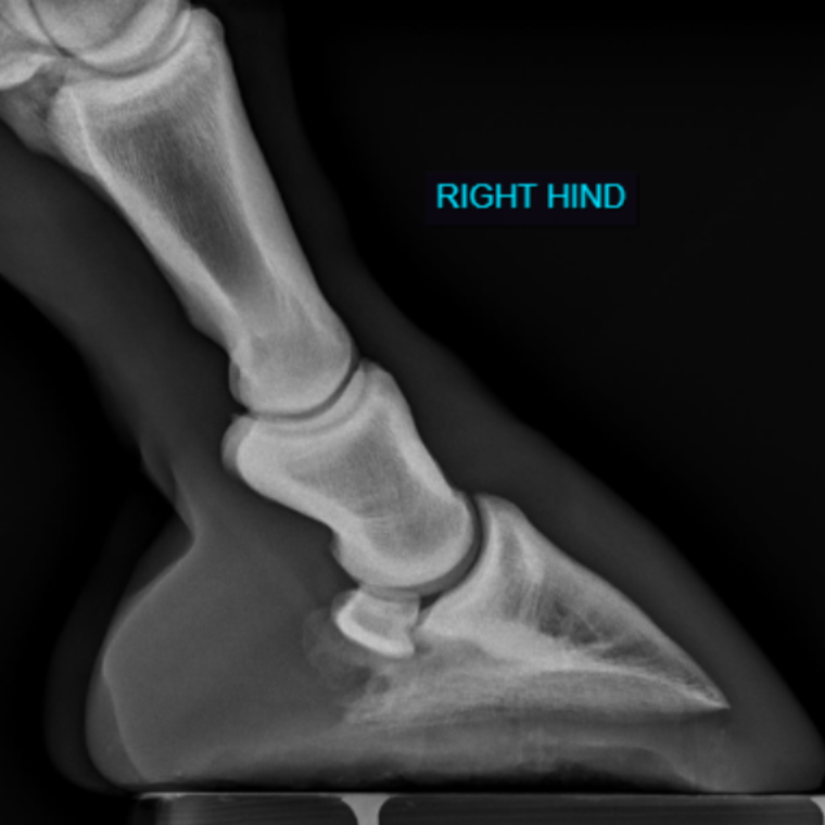
Right Hind
Radiograph of the right hind foot showing a negative plantar angle (heels of coffin bone are closer to the ground than the toe) and broken back alignment of the foot and pastern. This is a biomechanical challenge that needs to be addressed.
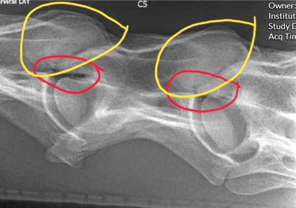
Radiograph showing enlarged cervical facets with roughening on the ventral (bottom) aspect of the joint circled in yellow. The area circled in red illustrates narrowing of the space where the nerves exit the vetebral body.

