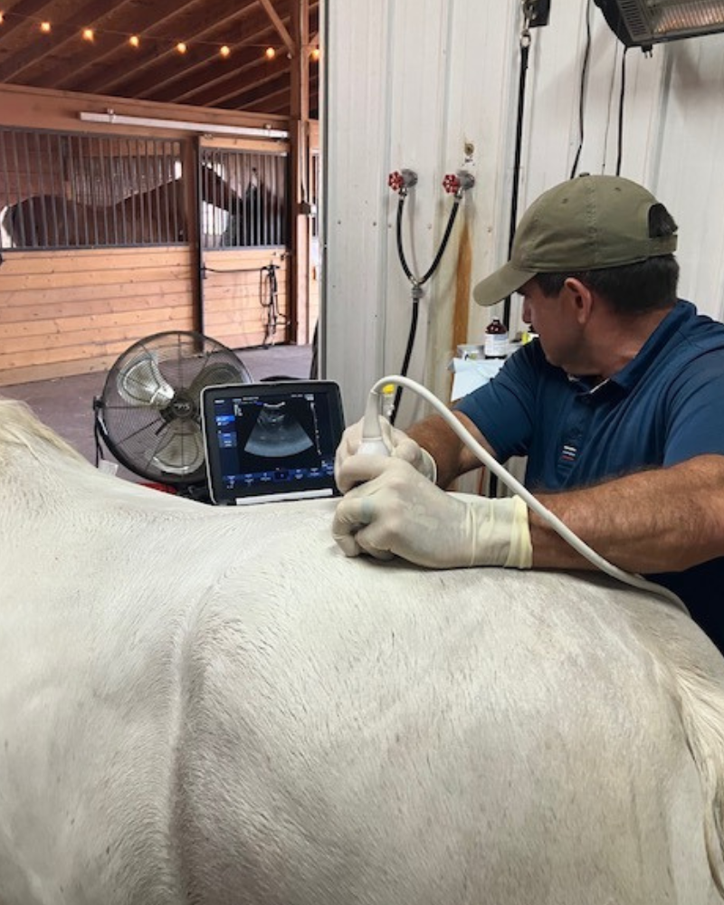
Digital ultrasound is used to evaluate tendons, ligaments, soft tissue, and bone surfaces. It clearly shows joint capsules, fluid quality, and helps assess damage for treatment decisions. Ultrasound-guided injections also enhance accuracy in treating joints and soft tissue injuries.
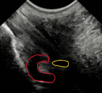
Ultrasound image shows excess fluid (distention or effusion) of the navicular bursa circled in red, and the coffin joint in yellow. Both of these structures would benefit from intra-articular therapy.

Ultrasound of Coffin Joint Navicular Bursa

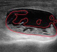
Ultrasound of Stifle
Ultrasound image of the medial stifle joint, which is what we typically treat when injecting stifles. The area circled in red shows synovitis and capsulitis (inflammation of the joint structures). This is a joint that is in definite need of intra-articular therapy.
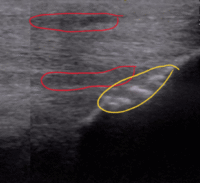
Ultrasound image shows suspensory branch inflammation with fiber pattern disruption circled in red, and avulsion of bone where the ligament inserts on the sesamoid bone. This would have to correlated with a physical exam to determine if it a source of lameness.

