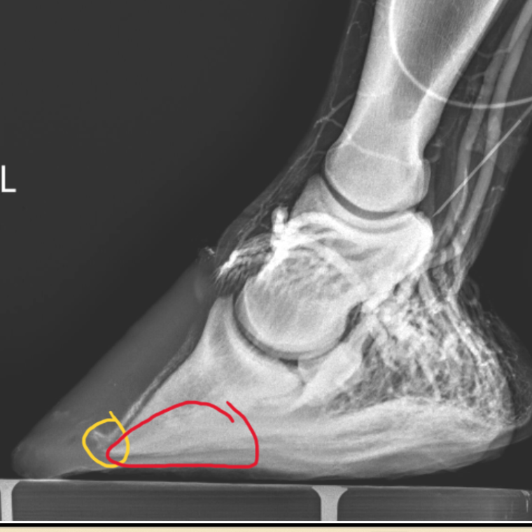
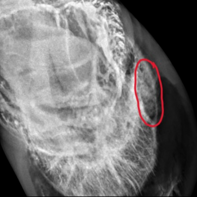
Venograms are used to evaluate the condition of blood vessels in the feet of laminitic horses. A tourniquet is applied at the fetlock, and a radiopaque dye (visible on the radiograph) is injected into a vein above the foot. Radiographs are then taken from multiple angles to visualize the vasculature. This process assists in determining the severity of the condition, appropriate treatment approaches, and prognosis. Additionally, venograms can help identify abscesses, foot infections, and keratomas by revealing interruptions in blood supply.
Venogram Lateral: This venogram of a laminitis case illustrating the complete lack of blood supply in the terminal arch (central part of the foot) and sole circled in red. The yellow circle identifies the circumflex vessels distorted and displaced proximally. This is a clear case showing the catastrophic damage that’s not fully appreciated on the plan radiograph.

Venogram Lateral

Venogram Infection
Venogram infection: This venogram shows disruption of the blood supply circled in red. This is an abscess that we were unable to find looking at the foot or on regular radiographs. We were able to drain the abscess through the wall and resolve the problem.
